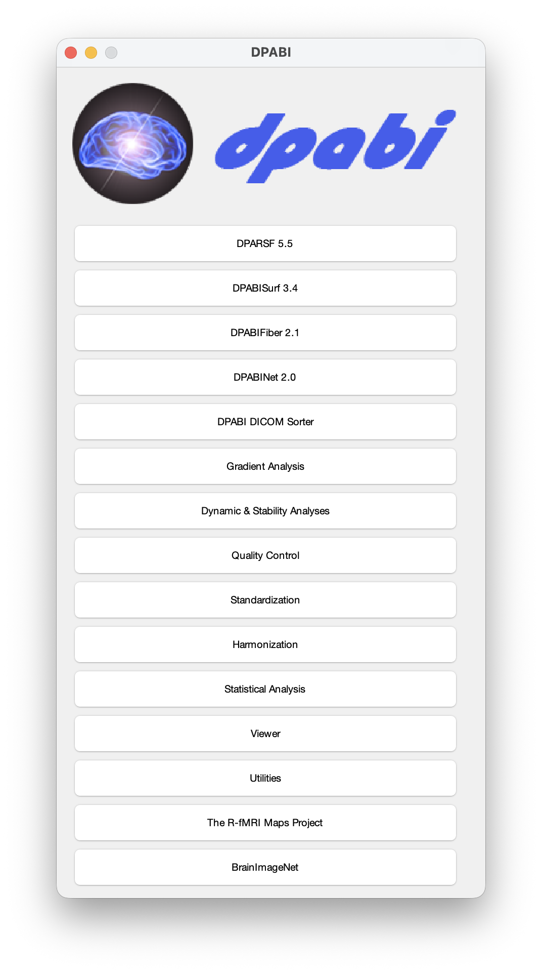
DPABI: a toolbox for Data Processing & Analysis for Brain Imaging
New features of DPABI_V10.0_260625 (download at http://rfmri.org/dpabi, please also update the dpabi, dpabifiber, qsiprep and qsirecon docker files by: docker pull cgyan/dpabi and so on):
1. DPABISurf 3.4. Multi Echo fMRI Data pipeline was included (based on Tedana). Please read the usage from Lesson 6 and Lesson 12 at https://edu.deepbrain.com/RFMRICourse (or this video: https://rfmri.org/share/DPABI_Data_Organization_20260705.mp4)
2. DPABINet 2.0. DPABI Structure-Function Coupling Analysis module was included. Please read the usage from Lesson 24 at https://edu.deepbrain.com/DPABINetAdvancedCourse.
3. DPABIFiber 2.1.
3.1. Convert freesurfer aparcaseg files to qsiprep ACPC space.
3.2. Convert FC files to qsiprep ACPC space for TWFC.
3.3. Fixed a bug in normalizing TDI maps in new DPABIFiber
4. DPABI Gradient Analysis module was included (based on Brainspace). Please read the usage from Lesson 31 at https://edu.deepbrain.com/RFMRICourse.
5. DPABI Harmonization. Harmonizing the brain images (.nii/.nii.gz/.gii/.mat) to remove site effects for big data in statistical analysis. Please read the usage from Lesson 30 at https://edu.deepbrain.com/RFMRICourse.
6. Added support for data from United Imaging's uMR Jupiter 5T. Please read the usage from Lesson 6 at https://edu.deepbrain.com/RFMRICourse (or this video: https://rfmri.org/share/DPABI_Data_Organization_20260705.mp4)
7. To use DPABI V9, you need to use the developing version of SPM: https://github.com/spm/spm or use this alternative one.
Tips:
1) For Linux or Mac OS, please start matlab from terminal in order to reach docker in DPABI (e.g., Linux: matlab; Mac: open /Applications/MATLAB_R2018a.app/).
2) Before running DPABISurf_Pipeline, you can test the docker environment by running DPABI->DPABISurf->Utilities->Volume-Surface Projector. If the file can be successfully projected to surface, then the software is all set.
New features of DPABI_V9.0_250415 (download at http://rfmri.org/dpabi, please also update the dpabi, freesurfer, dpabifiber, qsiprep and qsirecon docker files by: docker pull cgyan/dpabi and so on):
1. New Recommended Data Processing Workflow from DICOM Files
1.1 If your dataset includes DICOM files, the processing logic has been updated — you are recommended to now begin with DPABI DICOM Sorter. From there, you will proceed through the new DPABI Input Preparer, which automatically organizes various types of imaging data, including T1, T2, functional MRI, DWI, and field maps.
1.2. This new pipeline can automatically call DPARSF and DPABISurf in sequence. Subsequently, DPABISurf and DPABIFiber will handle all types of data — including various kinds of field maps — in an integrated and automated manner.
2. DPABISurf 3.3
2.1. Backend engine updated to fMRIPrep V24.1.1.
2.2. The Field Map button has been removed. However, if you use DPABI Input Preparer to organize your field map data, field map correction will be automatically integrated during processing.
3. DPABIFiber 2.0
3.1. Backend engine updated to QSIPrep and QSIRecon V1.0.0.
3.2. Field map correction is now automatically integrated if your data is organized via DPABI Input Preparer.
3.3. A new option has been added: you can define a custom reconstruction pipeline by creating {WorkingDir}/Custom.yaml.
4. DPARSF 5.5
4.1. In DPARSFA, the logic remains different from DPABISurf and DPABIFiber — you still need to manually click the Field Map button. Note: The unit of Echo Time has changed from milliseconds (ms) to seconds (s).
4.2. Fixed a bug related to Automask when calling Docker under Windows.
New features of DPABI_V8.2_240510 (download at http://rfmri.org/dpabi, please also update the dpabi and freesurfer docker file by: docker pull cgyan/dpabi and docker pull cgyan/freesurfer):
1. DPABISurf 3.2.
1.1. Back engine updated to fMRIPrep V23.2.2.
1.2. "Segment Subregions" now using Freesurfer 7.4.1. Thus solved the failure issues when segmenting subregions for some datasets.
1.3. DPABISurfSlurm is updated accordingly.
2. DPABI Harmonization.
2.1. Site Info is moved from subGUI to main GUI.
2.2. Add new functions for Create FileList and Config save and load.
2.3. Fixed a bug for skipping a lot of zero voxels.
3. DPABI_VIEW: added Yeo 2011 7 and 17 networks for atlas.
4. DPABI Dynamic & Stability Analyses: Fixed a bug in loading fsaverage5_hemi-R*.func.gii files.
5. DPARSF: Fixed a checking docker bug in windows for AutoMask.
6. Changed to webread in case under some internet issues, thanks to the suggestion of Roger.
New features of DPABI_V8.1_240101 (download at http://rfmri.org/dpabi):
1. DPABISurf 3.1.
DPABISurfSlurm Updated: High Performance Computing Version of DPABISurf was updated after processing 4021 subjects on a HPC. Please read here for how to use it: http://rfmri.org/DPABISurfSlurm
2. DPABI Harmonization module was updated, please also update ICVAE docker: docker pull cgyan/icvae.
New features of DPABI_V8.0_231111 (download at http://rfmri.org/dpabi, please also update the docker file by: docker pull cgyan/dpabi and docker pull cgyan/dpabifiber):
1. DPABI Harmonization module was released.
This module could be used to harmonizing the brain images (.nii/.nii.gz/.gii/.mat) to remove site effects for big data in statistical analysis. Please read our latest Reference: Wang, Y.W., Chen, X., Yan, C.G. (2023). Comprehensive evaluation of harmonization on functional brain imaging for multisite data-fusion. Neuroimage, 274, 120089, doi:10.1016/j.neuroimage.2023.120089.
Please see a tutorial for DPABI Harmonization Module here (in Chinese), please download the Demo Data for DPABI Harmonization Module from here.
2. DPABISurf 3.0.
2.1. DPABISurfSlurm: High Performance Computing Version of DPABISurf was released. Please read here for how to use it: http://rfmri.org/DPABISurfSlurm
2.2. Back engine updated to fMRIPrep V23.1.4, thus using FreeSurfer 7.3.2.
2.3. Segment subregions (Thalamus, Hippocampus, Amygdala and Brainstem) with FreeSurfer 7.3.2 was set to the default option now.
2.4. Added freesurfer atlases: {DPABISurf}/SurfTemplates/fsaverage5_lh_aparc_a2009s_annot.label.gii and fsaverage5_lh_aparc_annot.label.gii.
2.5. Could make use of DPABI Harmonization module.
3. DPABIFiber 1.1.
3.1. Back engine updated to QSIPrep V0.19.1.
3.2. Could make use of DPABI Harmonization module.
4. DPABINet 1.3.
4.1. Could make use of DPABI Harmonization module.
5. Fixed a bug in handling surface correlation analysis in y_Correlation_Image.m.
6. Fixed a bug of "Brace indexing is not supported for variables of this type" in DPABI_TDA.
New features of DPABI_V7.0_230110 (download at http://rfmri.org/dpabi, please also update the docker file by: docker pull cgyan/dpabi): 1. DPABIFiber 1.0.
DPABIFiber is a fiber tractography analysis toolbox based on diffusion-weighted imaging (DWI), evolved from DPABI/DPABISurf/DPABINet/DPARSF, as easy-to-use as DPABI/DPABISurf/DPABINet/DPARSF. DPABIFiber is based on QSIPrep (Cieslak et al., 2021), MRtrix3 (Tournier et al., 2019), AFQ (Yeatman et al., 2012), fMRIPprep (Esteban et al., 2019), FreeSurfer (Tustison et al., 2014), ANTs (Avants et al., 2009), FSL (Jenkinson et al., 2012), SPM12 (Ashburner, 2012), dcm2niix (Li et al., 2016), PALM (Winkler et al., 2014), GNU Parallel (Tange, 2011), MATLAB (The MathWorks Inc., Natick, MA, US), Docker (https://docker.com) and DPABI (Yan et al., 2016). DPABIFiber provides a user-friendly graphical user interface (GUI) for pipeline DWI preprocessing, fiber tractography reconstruction, tract-based spatial statistics (TBSS) (Smith et al., 2006), automating fiber-tract quantification (AFQ) (Yeatman et al., 2012), structural connectome matrix analyses, seed-based structural connectivity analyses, and tract-weighted functional connectivity (TW-FC) (Calamante et al., 2013), while requires no programming/scripting skills from the users. Please see more details at: http://rfmri.org/DPABIFiber
2. DPABISurf 2.0.
2.1. Back engine updated to fMRIPrep V22.1.1, thus using FreeSurfer 7.
2.2. Added new mode: Calculate in Native Space. The processed results in native space also fits better to the TWFC analyses with DPABIFiber Pipeline.
2.3. Could segment subregions (Thalamus, Hippocampus, Amygdala and Brainstem) with FreeSurfer 7.3.
2.4. Could use FastSurfer instead of FreeSurfer to perform surface construction.
2.5. Could add wildcard strings in Defining ROIs.
2.6. Could select ROI Indices for specific ROI files.
2.7. Could delete first time points if start with BIDS.
2.8. Could detect TR if start with BIDS or fmriprep.
3. DPARSF 5.4.
3.1. Could add wildcard strings in Defining ROIs.
3.2. Could select ROI Indices for specific ROI files.
4. Statistical Analysis. Added option of TFCE2D for TBSS and surface-based statistical analysis.
5. y_ReadRPI. Fixed a bug in calculating new origin when flipping axes.
New features of DPABI_V6.2_220915 (download at http://rfmri.org/dpabi, you do NOT need to update the docker file as compared to V6.1): 1. This is the last release of DPABI V6, still using freesurfer 6. DPABI V7 will have more changes.
2. DPABISurf 1.8:
2.1. Fixed a bug in displaying negative values in DPABISurf_VIEW.
2.2. Added area and curv in DPABISurf templates.
3. DPABINet 1.2:
3.1. Some weighted graph theoretical metrics calculations were changed (matrix normalization, use only non-negative functional connectivity etc.). The GTA results for weighted metrics (e.g., Eglob, betweenness, Lp) may be DIFFERENT from previous version.
4. DPABI_VIEW:
4.1. Now support rotating the image. Try More->Reorientation. Usually you only need to set Pitch, Roll and Yaw. You can also save it by More -> Save Reoriented Images.
4.2. Now support set the display color range of underlay. Set it whey you load a user-defined underlay. Also can try "User-Defined" in the drop-down menu.
5. Updated y_Meta_Image_CallR.m to handle surface file.
6. Fixed a bug in w_StatToP.m in handling r value.
New features of DPABI_V6.1_220101 (download at http://rfmri.org/dpabi, please also update the docker file by: docker pull cgyan/dpabi): 1. DPARSF V5.3.
1.1. Use 3dAutomask in AFNI to generate automasks if docker is installed.
2. DPABISurf V1.7.
2.1. fMRIPrep backend updated to version 20.2.5 (Long-Term Support).
2.2. If the TR info in NIfTI header is different from what specified by the user in DPABISurf_Pipeline GUI, then rewrite as user specified.
3. DPABINet V1.1
3.1. Fixed a bug in DPABINet_VIEW when displaying negative edges.
4. DPABI Input Preparer is here! You will love it! DPABI->Utilities->DPABI Input Preparer.
5. Fixed all GUIs displaying issues on Windows and Linux platforms.
6. Fixed a bug in ROI Signal Extractor.
1. DPABINet V1.0 released!
DPABINet is a toolbox for brain network and graph theoretical analyses, evolved from DPABI/DPABISurf/DPARSF, as easy-to-use as DPABI/DPABISurf/DPARSF. DPABINet is based on Brain Connectivity Toolbox (Rubinov and Sporns, 2010) (RRID:SCR_004841), FSLNets (http://fsl.fmrib.ox.ac.uk/fsl/fslwiki/FSLNets; RRID: SCR_002823), BrainNet Viewer (Xia et al., 2013) (RRID:SCR_009446), circos (Krzywinski et al., 2009) (RRID:SCR_018207), SPM (Ashburner, 2012) (RRID:SCR_007037), PALM (Winkler et al., 2016), MATLAB (The MathWorks Inc., Natick, MA, US) (RRID:SCR_001622), Docker (https://docker.com) (RRID:SCR_016445) and DPABI (Yan et al., 2016) (RRID:SCR_010501). DPABINet provides user-friendly graphical user interface (GUI) for Brain network construction, graph theoretical analyses, statistical analyses and results viewing, while requires no programming/scripting skills from the users.
2. DPABISurf V1.6.
2.1. Can use individual ROI definition files for each subject. The first line of the ROI definition file should be: 'Seed_ROI_List:'. An example SeedROI.txt:
Seed_ROI_List:
/Processing/fmriprep/sub-Sub001/func/sub-Sub001_task-rest_space-MNI152NLin2009cAsym_desc-aseg_dseg.nii.gz
/Processing/fmriprep/sub-Sub002/func/sub-Sub002_task-rest_space-MNI152NLin2009cAsym_desc-aseg_dseg.nii.gz
/Processing/fmriprep/sub-Sub003/func/sub-Sub003_task-rest_space-MNI152NLin2009cAsym_desc-aseg_dseg.nii.gz
...
2.2. Fixed a bug in dealing with multiple sessions of FieldMap.
2.3. Allow skipping subjects in TRInfo.tsv.
2.4. The surface for ReHo calculation were changed from pial to white.
3. DPARSF V5.2.
3.1. Users can threshold subjects with bad quality after reorienting.
3.2. Allow skipping subjects in TRInfo.tsv.
4. DICOM sorter added new layout and show demo.
5. A BIDS converter was added. DPABI->Utilities->DPABI BIDS Converter. Users can also deface the T1 Images before converting to BIDS. This module is good for sharing BIDS data, and is compatible with Brain Imaging Sharing Initiative: http://bisi.org.cn.
6. As suggested by Dr. Andrew Zalesky, the functional subcortical atlas "Tian2020_Subcortex_Atlas" was added. Ref: Tian, Y., Margulies, D.S., Breakspear, M., Zalesky, A. (2020). Topographic organization of the human subcortex unveiled with functional connectivity gradients. Nat Neurosci, 23(11), 1421-1432, doi:10.1038/s41593-020-00711-6.
7. A function get_components.m within the Network-based statistic (NBS) toolbox was added with Dr. Andrew Zalesky's permission. Ref: Zalesky, A., Fornito, A., Bullmore, E.T. (2010). Network-based statistic: identifying differences in brain networks. Neuroimage, 53(4), 1197-1207, doi:10.1016/j.neuroimage.2010.06.041.
8. Brain Connectivity Toolbox (BCT) was redistributed within DPABI with Dr. Mikail Rubinov's permission. Ref: Rubinov, M., Sporns, O. (2010). Complex network measures of brain connectivity: Uses and interpretations. Neuroimage, 52(3), 1059-1069, doi:S1053-8119(09)01074-X [pii] 10.1016/j.neuroimage.2009.10.003.
9. DPABI now is compatible with Dr. Mingrui Xia's latest version of BrainNet Viewer. Ref: Xia, M., Wang, J., He, Y. (2013). BrainNet Viewer: a network visualization tool for human brain connectomics. PLoS ONE, 8(7), e68910, doi:10.1371/journal.pone.0068910.
New features of DPABI_V5.1_201201 (download at http://rfmri.org/dpabi, please also update the docker file by: docker pull cgyan/dpabi): 1. DPABISurf V1.5.
1.1. fMRIPrep backend updated to version 20.2.1 (Long-Term Support).
1.2. Fixed a bug during processing data with multiple sessions.
1.4. DPABISurf_VIEW. Fixed a bug when setting p = 0.001, but still display 1. Fixed a bug to input index. Added save colorbar.
2. As discussed with Drs. Edmund Rolls and Marc Joliot, the AAL3 template was integrated into DPABI.
4. dcm2niix updated to version 2-November-2020 (v1.0.20201102)
New features of DPABI_V5.0_201001 (download at http://rfmri.org/dpabi, please also update the docker file by: docker pull cgyan/dpabi): 1. New Module: BrainImageNet included! With BrainImageNet, the model can classify the sex of a participant with brain structural imaging data from anybody and any scanner with about 95% accuracy. The model fine-tuned to Alzheimer’s Disease (AD) achieved 88.4% accuracy in leave-sites-out five-fold cross-validation on the ADNI dataset and 86.1% accuracy for a direct test on an unseen independent dataset (OASIS). When directly testing this AD classifier on brain images of unseen mild cognitive impairment (MCI) patients, 63.2% who finally converted into AD were predicted as AD, versus 22.1% who did not convert into AD were predicted as AD. Predicted scores of the AD classifier showed significant correlations with severity of illness. Users can utilize our models as bases for transfer learning. Please read more details at https://www.biorxiv.org/content/10.1101/2020.08.18.256594v2.
2. DPABISurf V1.4.
2.1. fMRIPrep backend updated to version 20.2.0 (Long-Term Support).
2.2. For surface based multiple comparison correction, the method of FDR correction was added to DPABISurf. See DPABISurf_VIEW->Cluster...->Apply FDR Correction.
2.3. For surface based multiple comparison correction, the method of Monte Carlo simulation was added to DPABISurf. See DPABISurf_VIEW->Cluster...->Apply FWE (Monte Carlo Simulation) Correction.
2.4. Pre-calculated Monte Carlo Simulation tables were added for most used situations. Please see {DPABI}/StatisticalAnalysis/DPABISurf_MonteCarlo/MonteCarloTable/. Users can index the minimum cluster area for an estimated smoothness and set at DPABISurf_VIEW->Cluster...->Set Cluster Size.
3. DPARSF V5.1.
3.1. Fixed a bug in applying slice timing information from DICOM files to DPARSF settings. This bug only affected DPARSF V5.0 while not setting slice timing information (using the default) in slice timing preprocessing. Automatically applying slice timing information from DICOM files missed the unit change for SPM.
4. Scrubbing default setting changed to FD_Jenkinson, 0.2mm, no time points before or after "bad" time points.
5. PALM updated to version alpha115.
6. dcm2niix updated to version 31-March-2020 (v1.0.20200331)
7. DICOM Sorter. Now DICOM Sorter can handle files with DICOM files mixed with other kind of files when setting suffix to none.
8. Fixed a bug of "Add Image" in ROI Signals Extractor.
9. y_ANCOVA1_Multcompare_Image now is compatible for .gii.
10. Docker File: MCR files extracted for easy use in Singularity.
New features of DPABI_V4.3_200401 (download at http://rfmri.org/dpabi, please also update the docker file by: docker pull cgyan/dpabi): This is a minor update to fix some bugs.
1. DPARSF V5.0. Fixed a bug specific in Windows OS when starting from .nii.gz files: bet fails.
2. DPABISurf V1.3. Fixed the errors of "--ignore-aroma-denoising-errors" and "--template-resampling-grid". These were caused by the input format changing in fmriprep V20.
New features of DPABI_V4.3_200301 (download at http://rfmri.org/dpabi, please also update the docker file by: docker pull cgyan/dpabi): 1. Stability Analysis module was added. You can calculate volume-based and surface-based stability from DPABI->Dynamic & Stability Analyses. You can read our recent work for the details of stability measure: Li, L., Lu, B., Yan, C.G. (2019). Stability of dynamic functional architecture differs between brain networks and states. Neuroimage, 116230, doi:10.1016/j.neuroimage.2019.116230.
2. Field map correction was added, both for DPARSF and DPABISurf. If you want to perform field map Correction, you need to arrange each subject's field map DICOM files in one directory, and then put them in "FieldMap" directory under the working directory. i.e.:';...
'{Working Directory}\FieldMap\PhaseDiffRaw\Subject001\xxxxx001.dcm';...
'{Working Directory}\FieldMap\PhaseDiffRaw\Subject001\xxxxx002.dcm';...
'...';...
'{Working Directory}\FieldMap\PhaseDiffRaw\Subject002\xxxxx001.dcm';...
'{Working Directory}\FieldMap\PhaseDiffRaw\Subject002\xxxxx002.dcm';...
'...';...
'{Working Directory}\FieldMap\Magnitude1Raw\Subject001\xxxxx001.dcm';...
'{Working Directory}\FieldMap\Magnitude1Raw\Subject001\xxxxx002.dcm';...
'...';...
'{Working Directory}\FieldMap\Magnitude1Raw\Subject002\xxxxx001.dcm';...
'{Working Directory}\FieldMap\Magnitude1Raw\Subject002\xxxxx002.dcm';...
'...';...
'...';...
Then you can click the button of “FieldMap” button to set field map correction parameters. In most cases, you can use the default “0” value to let the program read the parameters (e.g., echo times) from the DICOM files.
3. Check data organization function added. For the new users of DPARSF and DPABISurf, most of the errors were caused by data organization! Please use DPABI->Utilities->Check Data Organization to check your data organization before running DPARSF or DPABISurf. This program will lead you to organize your data correctly with prompting messages!
4. Slice timing information read from DICOM files. If you are starting with DICOM files, you no longer need to set the slice timing correction parameters. Just leave it as default (slice number: 0), then DPARSF or DPABISurf will read the parameters from DICOM files. This new feature thanks to Dr. Chris Rorden's new dcm2niiX program (version v1.0.20190902).
5. DPARSF V5.0 now is compatible with BIDS format. You can start with BIDS format data by checking checkbox “BIDS to DPARSF” and setting “Starting Directory Name” to “BIDS”.
6. DPABISurf V1.3. Check and re-run fmriprep failed subjects. If for any reason, the program failed fmriprep running in DPABISurf, you just need to re-run starting with the step “Preprocessing with fmriprep” and set the “Starting Directory Name” to “BIDS” in DPABISurf_Pipeline. Alternatively, you can run a single checking step from DPABI->DPABISurf->Utilities->Re-Run fmriprep Failed Subjects.
7. DPABI->Utilities->DICOM Sorter. Now DICOM Sorter will remove illegal characters for file names in DICOM sorter. E.g., a subject id of “Wang’#$#’s” will no longer cause a problem in sorting DICOM files.
8. y_Call_DPABISurf_VIEW_FromVolume.m and y_Call_DPABISurf_VIEW.m were added. Now you can use script to call DPABISurf_VIEW to generate surface maps (with higher quality now) in batch. Remember to add “close all” in the for loop to prevent too many windows.
New features of DPABI_V4.2_190919 (download at http://rfmri.org/dpabi, please also update the docker file by: docker pull cgyan/dpabi):
1. DPABISurf_V1.2_190919 updated.
1.1. A quality control module was added to DPABISurf. Now users can quality control surface reconstruction, EPI to T1 registration and T1 to MNI registration for all the subjects in one HTML file, respectively (based on fmriprep 1.5.0). For volume-based analysis, users can also generate group mask for DPABISurf, and exclude subjects by thresholding coverage and head motion.
1.2. DPABISurf now also output sulcus depth and volume in fsaverage and fsaverage5 spaces for statistical analysis.
1.3. In results organizer of DPABISurf, the redundant files would not be organized now. In addition, the fmriprep and freesurfer files were backed up, while excluding T1 image that may have privacy information such as face.
2. DPABI_VIEW has a new function "Surface View with DPABISurf_VIEW" now. This function will convert the files to fsaverage surface using freesurfer's mri_vol2surf command. Then the results were displayed by calling DPABISurf_VIEW to generate surface-based picture.
1. DPABISurf_V1.1_190725 updated.
1.1. New module for Surface-Based Temporal Dynamic Analysis (DPABI_TDA_Surf) was added. Dynamic regional indices (ALFF, fALFF, ReHo, Degree Centrality and Global Signal Correlation) and dynamic functional connectivity could be automatically calculated by one click through DPABI_TDA_Surf (with DPABISurf preprocessed data). The statistics maps (CV, Mean and SD) of the dynamic regional indices would also be generated by DPABI_TDA_Surf. A neuroimaging index which measures the concordance of the dynamic regional indices is incorporated into DPABI_TDA_Surf. Please see more details at: Yan, C.-G., Yang, Z., Colcombe, S.J., Zuo, X.-N., Milham, M.P., 2017. Concordance among indices of intrinsic brain function: insights from inter-individual variation and temporal dynamics. Sci Bull 62, 1572-1584.
1.2. The calculation of degree centrality now considers a vertex's correlation to both left and right hemispheres.
1.3. Standardization considers bilateral hemispheres.
1.4. Smooth function after Standardization was added.
1.5. If ICA-AROMA was chosen, no head motion realign parameters would be regressed out.
1.6. Fixed compatibility issues with old matlab versions.
1.7. The default surface-based smoothing kernel changed to 6mm instead of 10mm.
1.8. The DPABISurf results organizing function was added to the R-fMRI Maps Project.
1.9. Output an excel table for the volume of subcortical structures (calculated by freesurfer): {WorkingDir}/Results/AnatVolu/Anat_Segment_Volume.tsv.
1.10. DPABISurf_VIEW, the surface-based viewer now has a function to yoke between different viewers.
1.11. Docker updated basing on fMRIPrep 1.4.1. Besides pull from docker hub, the docker file can be also downloaded form baidu (extract code: enmn). 1.12. Besides the stand alone version of DPABI (with GUI), the compiled version of DPABISurf_run was also added to docker. Users can run DPABISurf_run with scripting. E.g.,
docker run -it --rm -v /My/FreeSurferLicense/Path/license.txt:/opt/freesurfer/license.txt -v /My/Data/Path:/data cgyan/dpabi /bin/bash
/opt/DPABI/DPABI_StandAlone/run_DPABISurf_run_StandAlone.sh ${MCRPath} /data/DPABISurf_Cfg.mat
2. DPARSF_V4.5_190725 updated.
2.1. For Linux or Mac OS, if FSL is not installed, then DPARSF will call FSL's bet in dpabi docker.
Tips for Linux or Mac OS: please start matlab from terminal in order to reach docker in DPABI (e.g., Linux: matlab; Mac: open /Applications/MATLAB_R2018a.app/).
1. DPABISurf was released! DPABISurf is a surface-based resting-state fMRI data analysis toolbox evolved from DPABI/DPARSF, as easy-to-use as DPABI/DPARSF. DPABISurf is based on fMRIPprep 1.3.0.post3 (Esteban et al., 2018)(RRID:SCR_016216), and based on FreeSurfer 6.0.1 (Dale et al., 1999)(RRID:SCR_001847), ANTs 2.2.0 (Avants et al., 2008)(RRID:SCR_004757), FSL 5.0.9 (Jenkinson et al., 2002)(RRID:SCR_002823), AFNI 20160207 (Cox, 1996)(RRID:SCR_005927), SPM12 (Ashburner, 2012)(RRID:SCR_007037), PALM alpha112 (Winkler et al., 2016), GNU Parallel (Tange, 2011), MATLAB (The MathWorks Inc., Natick, MA, US) (RRID:SCR_001622), Docker (https://docker.com) (RRID:SCR_016445), and DPABI V4.0 (Yan et al., 2016)(RRID:SCR_010501). DPABISurf provides user-friendly graphical user interface (GUI) for pipeline surface-based preprocessing, statistical analyses and results viewing, while requires no programming/scripting skills from the users. The DPABISurf pipeline first converts the user specified data into BIDS format (Gorgolewski et al., 2016), and then calls fMRIPprep 1.3.0.post3 docker to preprocess the structural and functional MRI data, which integrates FreeSurfer, ANTs, FSL and AFNI. With fMRIPprep, the data is processed into FreeSurfer fsaverage5 surface space and MNI volume space. DPABISurf further performs nuisance covariates regression (including ICA-AROMA) on the surface-based data (volume-based data is processed as well), and then calculate the commonly used R-fMRI metrics: amplitude of low frequency fluctuation (ALFF) (Zang et al., 2007), fractional ALFF (Zou et al., 2008), regional homogeneity (Zang et al., 2004), degree centrality (Zuo and Xing, 2014), and seed-based functional connectivity. DPABISurf also performs surface-based smoothing by calling FreeSurfer’s mri_surf2surf command. These processed metrics then enters surfaced-based statistical analyses within DPABISurf, which could perform surfaced-based permutation test with TFCE by integrating PALM. Finally, the corrected results could be viewed by the convenient surface viewer DPABISurf_VIEW, which is derived from spm_mesh_render.m. Please see details at http://rfmri.org/DPABISurf. 2. DICOM Sorter: In case PatientID is not defined, use PatientName.FamilyName instead.
1. Added a prompt of "Congratulations, the running of DPARSFA is done!!! :)" when DPARSF finishes its processing.
2. Added a new atlas (Schaefer2018_400Parcels_7Networks_order_FSLMNI152_1mm.nii) to the V4 parameters. Please see more details at Schaefer, A., Kong, R., Gordon, E.M., Laumann, T.O., Zuo, X.N., Holmes, A.J., Eickhoff, S.B., Yeo, B.T.T., 2017. Local-Global Parcellation of the Human Cerebral Cortex from Intrinsic Functional Connectivity MRI. Cereb Cortex, 1-20.
3. The dcm2nii has been updated to the latest version in courtesy of Dr. Chris Rorden. See: Li, X., Morgan, P.S., Ashburner, J., Smith, J., Rorden, C., 2016. The first step for neuroimaging data analysis: DICOM to NIfTI conversion. J Neurosci Methods 264, 47-56.
4. As there were some parallel computing issues in calling outside command, the callings were no longer using parallel computing (i.e., downgrade from parfor to for). These includes the callings of dcm2nii and bet.
5. Flexibility for concordance was added to the module of Temporal Dynamic Analysis (DPABI_TDA). Users can freely calculate the concordance of any combinations of ALFF, fALFF, ReHo, Degree Centrality, Global Signal Correlation and VMHC.
6. Fixed some compatibility bugs with higher versions of MATLAB. For example, Time Course error in DPABI_VIEW; uimenu parent problem when calling monkey/rat module; errors regard generating pictures for checking normalization in DPARSFA.
7. Tips for calling "bet": You should start matlab from terminal (e.g., Linux: matlab; Mac: open /Applications/MATLAB_R2018a.app/). If you installed FSL5.0, you may also need to run this: source /usr/share/fsl/5.0/etc/fslconf/fsl.sh. In addition, in some Linux versions, you may need to start matlab in this way: LD_PRELOAD="/usr/lib/x86_64-linux-gnu/libstdc++.so.6" matlab.
1. New module for Temporal Dynamic Analysis (DPABI_TDA) was added. Dynamic regional indices (ALFF, fALFF, ReHo, Degree Centrality, Global Signal Correlation and VMHC) and dynamic functional connectivity could be automatically calculated by one click through DPABI_TDA (with DPARSF preprocessed data). The statistics maps (CV, Mean and SD) of the dynamic regional indices would also be generated by DPABI_TDA. A new neuroimaging index which measures the concordance of the dynamic regional indices is incorporated into DPABI_TDA. Please see more details at: Yan CG, Yang Z, Colcombe S, Zuo XN, Milham MP (2017) Concordance among indices of intrinsic brain function: insights from inter-individual variation and temporal dynamics. Science Bulletin. In press.
2. The default setting of permutation test with PALM was changed to two-tailed test. According to our recent study, permutation test with Threshold-Free Cluster Enhancement (TFCE) reaches the best balance between family-wise error rate (under 5%) and test-retest reliability / replicability, thus outperforms the other multiple comparison correction strategies. Please consider use it. Chen, X., Lu, B., Yan, C.G.*, 2018. Reproducibility of R-fMRI metrics on the impact of different strategies for multiple comparison correction and sample sizes. Hum Brain Mapp 39, 300-318.
3. Statistical Analysis Module. Also output effect size maps: Cohen’s f2 maps.
4. Statistical Analysis Module. Added a function for image-based meta analysis: y_Meta_Image_CallR.m. This functional is calling R package ‘metansue’, please install ‘metansue’ and ‘R.matlab’ first.
5. Statistical Analysis Module. Mixed Effect Analysis: Fixed a bug of OtherCovariates and CovVolume.
6. y_ExtractROISignal.m. Remove the voxels with “NaN” values.
7. DPARSF_V4.3_171210: If the file name before realignment is initialed with 'r', then move the 'rr*' files for the next step.
1. DPARSFA V4 Parameters (Default Parameters, also for The R-fMRI Maps Project). For ROI signals extraction, the Power 264 ROIs were added as the 1570~1833 ROIs. (Power_Neuron_264ROIs_Radius5_Mask.nii was added to {DPABI}/Templates/)
2. DPARSF Advanced Edition: Re-run with global signal regression (DPARSFA_RerunWithGSR). Fixed a bug when “Remove first X time points” was defined, the number of time points will be adjusted accordingly.
3. DPARSF. Add a “Clear All” button in the ROI List GUI for defining ROIs.
4. Statistical Analysis: Fixed a bug when other covariates were defined in y_MixedEffectsAnalysis_Image.m.
5. Added Yeo2011_7Networks_Colormap_ForDPABI.mat and Yeo2011_17Networks_Colormap_ForDPABI.mat to {DPABI}/Templates, for visualizing Yeo and Buckner 7 or 17 networks in DPABI Viewer.
6. Brainnetome Atlas was added to {DPABI}/Templates: BrainnetomeAtlas_BNA_MPM_thr25_1.25mm.nii.gz and BrainnetomeAtlas_BNA_subregions.xlsx. This is the Maximum Probabilistic Map (MPM) of Brainnetome Atlas, including 246 subregions (210 cortical and 36 subcortical subregions). Citation: Fan, L., Li, H., Zhuo, J., Zhang, Y., Wang, J., Chen, L., Yang, Z., Chu, C., Xie, S., Laird, A.R., Fox, P.T., Eickhoff, S.B., Yu, C., Jiang, T., 2016. The Human Brainnetome Atlas: A New Brain Atlas Based on Connectional Architecture. Cereb Cortex 26, 3508-3526.
7. DPABI Viewer. Added a button “Apply a Mask for Additionally Thresholding” under “Cluster”. This function is used for viewing permutation test results from statistical analysis. Please see http://wiki.rfmri.org/PermutationTest for detailed description for performing permutation test and visualizing the results.
1. DPARSF V4.2_161201 released.
1.1. To let the users be more aware what kind of templates they are using, SPM Templates were included under {DPABI}/Templates/ now. If you want to USE YOUR OWN TEMPLATES, please replace the corresponding ones under this directory instead of replacing those under SPM. For example: if you are using normalize by New Segment + DARTEL, please replace {DPABI}/Templates/SPMTemplates/tpm/TPM.nii; If you are using normalize by using EPI template, please replace {DPABI}/Templates/SPMTemplates/toolbox/OldNorm/EPI.nii; If you are using normalize by using T1 image unified segmentation, please replace {DPABI}/Templates/SPMTemplates/toolbox/OldSeg/grey.nii, white.nii, and csf.nii.
1.2. DPARSF Windows version. Previously need to run as administrator to get results both with and without global signal regression (GSR). Now such limitation is removed (change mklink to copyfile).
1.3. DPARSF Advanced Edition Preprocessing for Task fMRI data: For nuisance regression, the option of “Add mean back” is now default. The mean will be added back to the residual after nuisance regression. This is useful for circumstances of ICA or task-based analysis.
1.4. DPARSFA V4 Parameters (Default Parameters, also for The R-fMRI Maps Project). For ROI signals extraction, the global signal (BrainMask_05_91x109x91.img) was added as the 1569th ROI.
2. Statistical Analysis.
2.1. Given the recent concerns regarding multiple comparison correction, especially after Eklund et al. 2016 PNAS paper, We have included permutation test in the Statistical Analysis module. The permutation test was achieved by integrating PALM package, with the kind permission by Dr. Anderson M. Winkler. Please click “Permutation test (PALM)” button on the Statistical Analysis panel to use it. Please read http://fsl.fmrib.ox.ac.uk/fsl/fslwiki/PALM/UserGuide for the details of PALM. Please cite Winkler, A.M., Ridgway, G.R., Douaud, G., Nichols, T.E., Smith, S.M., 2016. Faster permutation inference in brain imaging. Neuroimage 141, 502-516 if you used it. 2.2. AlphaSim. For the so-called “bug” — the edge effects within the mask (apply mask and then smooth), DPABI doesn’t have such an issue since DPABI_V1.2_141101. On September 17, 2014, Dr. Katharina Wittfeld reported a bug with AlphaSim for small masks in combination with high smoothness: applying mask before the Gauss filter while the Gauss filter will blur the boundaries of the masked region which will cause problems later (http://rfmri.org/content/alphasim-problem-critical-bug). The code was revised to smooth the whole bounding box first and apply mask later, and the code was distributed with DPABI_V1.2_141101. The estimation of mean and standard deviation is within the whole bounding box as well, which we believe is better than estimating in a small mask (estimation of mean and standard deviation within a small mask might be inaccurate). In addition, Dr. Robert W Cox noted: "Simulations were also repeated with the now infamously "buggy" version of 3dClustSim: the effect of the bug on FPRs was minimal (of order a few percent)." http://biorxiv.org/content/early/2016/07/26/065862. 2.3. AlphaSim. The previous GUI version can only output simulation results for corner connection (Nearest Neighbor 26). Now we output 3 versions: face (NN6), edge (NN18) and corner (NN26) connection.
2.4. Mixed Effect Analysis (within-subject factor by between-subject factor) was added to the Statistical Analysis module. The order of the group images should be: Group1Condition1; Group1Condition2; Group2Condition1; Group2Condition2. You will get: *_ConditionEffect_T.nii - the T values of condition differences (corresponding to the first condition minus the second condition) (WithinSubjectFactor); *_Interaction_F.nii - the F values of interaction (BetweenSubjectFactor by WithinSubjectFactor); *_Group_TwoT.nii - the T values of group differences (corresponding to the first group minus the second group), of note, the two conditions will be averaged first for each subject for Group_TwoT analysis. (BetweenSubjectFactor).
3. A new ICC version based on R was included (y_ICC_Image_LMM_CallR.m), as the previous one (y_ICC_Image_LMM.m) fails to converge in many cases. y_ICC_Image_LMM_CallR.m is based on the R code written by Dr. Ting Xu (R_Cal_ICC.R). Of note, as this one needs the users to configure R environment (install.packages("nlme") and install.packages("R.matlab”)), the DPABI_ICC_TOOL (DPABI->Utilities->Test-Retest Reliability: ICC) GUI is still using y_ICC_Image_LMM.m. The new version (y_ICC_Image_LMM_CallR.m) should be used in command line.
4. DPABI Results Organizer and Intermediate Files Organizer (under “The R-fMRI Maps Project”): revised the parfor loop to prevent errors in case with too many files.
5. y_T1ImgAverager.m (DPABI->Utilities->T1 Images Averager): All the NaN voxels (in some cases) are set to zero now. Previously, the NaN voxels after averaging can induce trouble in bet.
6. MATLAB 2016b compatible.
New features of DPABI_V2.1_160415:
1. DPARSF V4.1_160415 released.
1.1. Fixed a bug in DPARSF Basic Edition. The bug is that the white matter signal is always removed in nuisance regression (only exist in the Basic Edition). Thanks to the report of Liviu Badea.
1.2. DPARSF Advanced Edition: Add an option of “Add mean back” for nuisance regression. The mean will be added back to the residual after nuisance regression. This is useful for circumstances of ICA or task-based analysis.
1.3. DPARSF Advanced Edition: Re-run with global signal regression (DPARSFA_RerunWithGSR). Fixed a bug when “Remove first X time points” was defined, the number of time points will be adjusted accordingly now. Thanks to the report of Hua-Sheng Liu.
1.4. DPARSF Advanced Edition: Add a slice timing batch mode for MultiBand data. Users could specify a text timing file for a given participant in SliceOrderInfo.tsv. Please see http://rfmri.org/SliceTiming for more details. 2. DPABI Results Organizer (under “The R-fMRI Maps Project”): also save the text version of ROI signals.
3. Dual Regression added (under Utilities). Define a map, then regress the map on the 4D data for a participant, thus get a time series. Variance-normalize the time series, and then regress on the 4D data, thus get the dual regression map.
4. DPABI Image Calculator (under “Utilities”). Syntax changed, now includes: g1.*To4D((i1>2.3),100) Make a mask (threshold at 2.3 on i1) and then apply to each image in group 1 (group 1 has 100 images).
5. Donsenbach 160 ROIs were merged into a single mask file (Dosenbach_Science_160ROIs_Radius5_Mask.nii).
6. Fixed a bug in y_GroupAnalysis_Image: CovVolume read error.
7. Add "MultiSelect" Mode when user selected "Add Image" for several GUIs.
8. DPABI Image Calculator (under Utilities): Fixed a bug when removing image or directory. Now, when you remove the item, the identifier will be re-ordered.
9. DPABI Viewer: Added a colorbar mode for DPABI_VIEW, when you add "+" or "-" flag at the end in "Add Overlay's Colorbar" entry, DPABI_VIEW will use full colormap to display the overlay.
New features of DPABI_V2.0_151201 (together with DPARSF_V4.0_151201):
1. Compatible with MATLAB 2014b and later versions.
2. Process the data both with and without global signal regression (GSR). Check “Nuisance regressors setting” -> “Both with & without GSR”. Alternatively, you can call DPARSFA_RerunWithGSR.m. E.g., DPARSFA_RerunWithGSR(DPARSFACfg.mat); where DPARSFACfg.mat stores the previous parameters without GSR.
3. The processing steps are affixed to Results directories. The R-fMRI calculation parameters are also written to the header of the result files.
4. V4 processing parameter template is added. No smoothing before R-fMRI measure calculation (except for VMHC). This is used for comparing across studies and accumulate processed data.
5. DPABI Statistical Analysis. Add multiple comparison test after ANOVA, e.g., 'tukey-kramer' or 'hsd', 'lsd', 'dunn-sidak', 'bonferroni’ or ‘scheffe' procedures.
6. DPABI_VIEW: compatible with BrainNet Viewer 1.5.
7. Fixed a "File too small" bug when .hdr/.img files are used.
8. Fixed a bug in y_Standardize.m: error when multiple files are defined.
9. Fixed a bug in DPABI Image Calculator: error in standard deviation calculation along the 4th dimension.
10. Results Organizer module: with this module, the users could organize the intermediate files for future processing with DPABI. In addition, the results could be organized for future use, and to be accumulated for the future R-fMRI maps project.
New features of DPABI_V1.3_150710:
1. SPM12 Compatible.
2. DPARSF_V3.2_150710 released.
3. DPARSF for Rat data released.
The Rat module is based on a Rat T2 template generated by Dr. Adam J. Schwarz et al. Please cite this paper when appropriate: Schwarz, A.J., Danckaert, A., Reese, T., Gozzi, A., Paxinos, G., Watson, C., Merlo-Pich, E.V., Bifone, A., 2006. A stereotaxic MRI template set for the rat brain with tissue class distribution maps and co-registered anatomical atlas: application to pharmacological MRI. Neuroimage 32, 538-550. (A T1 template was included as well. It's generated by normalizing 50 rats (two scans at PND45 or PND60) to that T2 template and then averaging (by Dr. Chao-Gan Yan)).
4. Fixed a bug in generating Voxel Specific Head Motion: missing gmdmp.
5. Fixed a bug in Group Analysis: when CovVolume is not defined.
6. Fixed a bug when calling BrainNet Viewer in DPABI_VIEW.
7. Fixed a bug in Standardization: ‘/‘ is not defined in Windows.
8. Fixed a bug in Image Calculator: output will be split as the input files when calculating group images.
New features of DPABI_V1.2_141101:
1. DPARSF V3.1 Basic Edition: Fixed a bug of missing DPARSF_run.
2. DPARSF V3.1: Fixed a bug that can not find ROI templates.
3. DICOM Sorter: Fixed a bug - “Add All” button doesn’t work.
4. DPABI Viewer: Fixed a bug when execute GRF or AlphaSim correction.
5. DPABI ROI Signal Extractor: New module added to DPABI->Utilities.
7. Test-Retest Reliability: Intraclass Correlation Coefficient (ICC). New module added to DPABI->Utilities. Three ICC algorithms are supported: ANOVA Model, ReML Model and Linear Mixed Models. The algorithms are based on Dr. Xi-Nian Zuo and his colleague’s work. Please cite Dr. Zuo’s work as detailed in each function.
New features of DPABI_V1.1_140827:
1. New modules in DPABI Utilities:
1.1. Multiple T1 Images Averager. If you have multiple T1 runs for each subject, this module will coregister them and make a mean T1 image to put to "T1Img" for following analyses.
2. Bugs Fixed.
2.1. DPABI Viewer: Cluster report doesn't work correctly after GRF correction. Thanks for Vincent's report! 3. DPABI now can check the latest version and pop up a notice.
DPABI includes the following components.
1. DPARSF 3.0 Advanced Edition.
New features in DPARSF 3.0 Advanced Edition.
1.1. Quality control. Integrated GUI for QCing the functional and structural images, users can give ratings and comments during the step of interactive reorientation.
1.2. Automask generation. For checking EPI coverage and generating group mask, the automasks (as in AFNI) will be generated based on EPI images.
1.3. Brain extraction (Skullstrip). This step can improve the coregistration between functional and structural images. Most registration issues of previous DPARSF versions can be solved by including this step. For Linux and Mac users: Need to install FSL. For Windows users: Thanks to Chris Rorden's compiled version of bet in MRIcroN, the modified version can work on NIfTI images directly.
1.4. Nuisance Regression. 1) Masks can be generated based on segmentation or SPM apriori masks; 2) Methods can be mean or CompCor [Note: for CompCor, detrend (demean) and variance normalization will be applied before PCA, according to (Behzadi et al., 2007)]; 3) Global Signal can be extracted based on Automasks.
2. DPARSF 3.0 Basic Edition.
2.1. DPARSF Basic Edition now is using the engine of DPARSF Advanced Edition.
2.2. Nuisance Regression (in MNI space) is placed before filtering, according to (Hallquist et al., 2013).
3. DPARSF for Monkey data.
3.1. The monkey module is based on Rhesus Macaque Atlases for functional and structural imaging studies generated by Wisconsin ADRC Imaging Core. Please cite their papers when appropriate: (McLaren et al., 2010; McLaren et al., 2009).
3.2. Of note, the origin of monkey atlas is different from human MNI atlas. Please make sure the correct origins are set at the steps of "reorienting Fun*" and "reorienting T1*".
4. Preprocessing for task fMRI.
Task fMRI data can be preprocessed via DPABI-DPARSF.
5. VBM.
VBM analyses can be performed via DPABI-DPARSF.
6. Quality Control.
6.1. QC Raw T1 images.
6.2. QC Raw functional images.
6.3. QC normalization effects. 1) QC on the pictures for checking spatial normalization. 2) Dynamically checking normalized T1, gray matter and functional images.
6.4. Thresholding QC scores and removing un-qualified subjects.
6.5. Generating Group masks based on normalized Automasks of each subject.
6.6. Thresholding EPI coverage.
6.7. Head motion report.
6.8. Thresholding head motion.
7. Standardization. Perform the following standardization according to (Yan et al., 2013).
7.1. Mean Regression
7.2. Mean Regression & SD Division
7.3. Mean Regression & Log SD Regression
7.4. Z - Standardization
7.5. Mean Division
7.6. Mean Subtraction
7.7. Median-IQR Standardization
7.8. Rank
7.9. Quantile Standardization
7.10. Gaussian Fit
8. Statistical Analysis.
Smoothness estimation based on the 4D residual is built in regression function – smoothness is written to the NIfTI headers automatically. For AlphaSim and GRF multiple comparison correction, only using smooth kernel applied in preprocessing is NOT sufficient, please use the estimated smoothness instead.
9. Viewer.
The DPABI_VIEW is based on spm_orthviews, but powered with convenient functions. Please try it out!
10. Utilities.
Utilities including DICOM Sorter, Image Calculator and Image Reslicer.
References
Behzadi, Y., Restom, K., Liau, J., Liu, T.T., 2007. A component based noise correction method (CompCor) for BOLD and perfusion based fMRI. Neuroimage 37, 90-101.
Hallquist, M.N., Hwang, K., Luna, B., 2013. The nuisance of nuisance regression: spectral misspecification in a common approach to resting-state fMRI preprocessing reintroduces noise and obscures functional connectivity. Neuroimage 82, 208-225.
McLaren, D.G., Kosmatka, K.J., Kastman, E.K., Bendlin, B.B., Johnson, S.C., 2010. Rhesus macaque brain morphometry: a methodological comparison of voxel-wise approaches. Methods 50, 157-165.
McLaren, D.G., Kosmatka, K.J., Oakes, T.R., Kroenke, C.D., Kohama, S.G., Matochik, J.A., Ingram, D.K., Johnson, S.C., 2009. A population-average MRI-based atlas collection of the rhesus macaque. Neuroimage 45, 52-59.
Yan, C.G., Craddock, R.C., Zuo, X.N., Zang, Y.F., Milham, M.P., 2013. Standardizing the intrinsic brain: towards robust measurement of inter-individual variation in 1000 functional connectomes. Neuroimage 80, 246-262.
Old Versions: V1.0 V1.1 V1.2 V1.3 V2.0 V2.1 V2.2 V2.3 V3.0 V3.1 V4.0 V4.1 V4.2 V4.3 V5.0 V5.1 V6.0 V6.1 V6.2 V7.0 V8.0 V8.1 V8.2 V9.0

您好!
您好!
Z standardization for ReHo and Degree Centrality
Dr. Yan,
Are the Z transforms for ReHo and Degree Centrality Fisher Z transforms, or are they some other Z transform? Additionally is there a citation or somewhere in the code where you could point where I could understand exactly how the values are being standardized? Thanks!
-Bryson
Yan, C.G., Craddock, R.C.,
Yan, C.G., Craddock, R.C., Zuo, X.N., Zang, Y.F., Milham, M.P., 2013. Standardizing the intrinsic brain: towards robust measurement of inter-individual variation in 1000 functional connectomes. Neuroimage 80, 246-262.
These Z transformations are minus brain mean and divide by brain STD.
Only VMHC is Fisher's Z transformed.
Multiple Seed Regions/DPARSF
Dear Dr. Yan
Would it possible to designate more than one ROI at one time?
What does the output files look like?
Cheers
Larry
Multiple Seed Regions/DPARSF - Fixed
It worked.
Thanks
Covariate warning
Hi all,
I am using DPARSFA 4.3, and during the covariate stage, I see the message:
Item 'Covariates', field 'val': Size mismatch (required [1 Inf], present [0 0]).
For each image. The program runs ok, and the covariates seem to have been applied. My covariates are: polynomial trend 2, Firston 24, WM and CSF (from SPM priors), and Compcor.
The results seem to be about the same as in prior work I've done using DPARSFA/DPABI. Is this a warning that can be ignored?
Thanks,
Matt
Hi Matt,
Hi Matt,
Could you post more warning information? Seems you can just ignore it.
Best,
Chao-Gan
local maxima/sub peaks in cluster needed
Dear all,
I did resting state analysis with dparsf advanced /DPABI (V3.0_171210) and got a big cluster for stronger connectivity with the IPL.
When I look at it with the Viever I can see that it covers several brain regions, but I can only read out one peak of the cluster with "Find peak in this cluster.How can I read out local maxima/subpeaks of different brain areas?
Thanks in advance,
Janine
We didn't write find local
We didn't write find local maxima/subpeaks.
As for now, you can enhance the threshold until it seperates. Then you can find the local maxima/subpeaks.
Thank you for your reply.
Thank you for your reply.
We already tried the threshold-enhancing method, but wanted to know, if there is a better way.
So, it is not possible to change something in the code of y_ClusterReport.m
to not only write out the amount of voxels for each subcluster, but also the peak of the reported subclusters?
Using DPABI Viewer
Hi Professor Chao-Gan,
First I want to thank you for developing this software. So far, I've been able to successfully view statistical maps, GFR corrected (voxel p<0.0214, cluster p <0.1 two-tailed). However, I would like to show a FC map one before and one after intervention as well. Is there a way to show an average FC map for all participants?
Or would there be a better program to visualize this? I've been trying to use BrainNetViewer with DPABI but was unable to do so. Should it work if the BrainNetViewer file is under the general DPABI folder, or should it be in a specific subfolder?
Please note that I'm very new to RS-fMRI analysis so apologies if my questions have already been answered.
Thank you so much,
Claire
Questions about CWAS
Dear Dr. Yan,
Thank you for developing such awesome software. I am a big fan of DPABI. Recently, I used y_CWAS.m to perform the CWAS analysis.
[p_Brain, F_Brain, Header] = y_CWAS(DataUpDir, SubID, AResultFilename, AMaskFilename, Regressor, iter, IsNeedDetrend, Band, TR)
I am a little confused of "Regressor". I know CWAS is an approach of identifying the pattern of connectome that relates to the clinical variable.
So, is this "Regressor" my clinical variable or the covariates (e.g. age and gender)?
If I input the clinical variable as "Regressor", where can I input the covariates (e.g. age and gender)?
Looking forward to your reply. Thank you so much. I really appreciate.
Best,
Clinical Variable, no
Clinical Variable, no covariates.
I haven't maintain this module for a long time. Thus you may need to check Shehzad's paper and see if you need to modify the codes.
load data
Please go over http://rfmri
Please go over http://rfmri.org/Course.
我想请教一些关于版本的问题
我刚接触这个软件,关于matlab版本以及dpabi、spm版本的问题不太了解,在运行的时候老是会有许多的错误,在网页中找不到manual的位置,有热心的朋友能帮忙给出一个可以用的matlab、dpabi、spm适配的版本么,谢谢
MATLAB 2015 or later. SPM12.
MATLAB 2015 or later. SPM12. Latest DPABI.
严老师:
严老师:
严老师:
严老师:
严老师:
严老师:
你是什么机器?新版DPABI更新了DICOM to
你是什么机器?新版DPABI更新了DICOM to NIfTI的组件。不过一般应该是没问题的。
DPABI install for linux some questions
Dear teacher: I come from Department of Psychiatry The 1st Affiliated Hospital of Kunming Medical University.After we completed the installation according to the tutorial, the next error occurred, which has not been resolved, please ask the teacher to help solve the next problem
Saving /home/labuser/dpabi/T1Img/Sub_003/ot1mpragesag.nii
Cropping NIfTI/Analyze image /home/labuser/dpabi/T1Img/Sub_003/ot1mpragesag.nii
Saving /home/labuser/dpabi/T1Img/Sub_003/cot1mpragesag.nii
Converting T1 Images:Sub_003 OK
Starting parallel pool (parpool) using the 'local' profile ...
connected to 12 workers.
Removing First 10 Time Points: Sub_001 OK
Removing First 10 Time Points: Sub_002 OK
Removing First 10 Time Points: Sub_003 OK
'Error in Removing First 10Time Points: Sub_001'
'Error in Removing First 10Time Points: Sub_002'
'Error in Removing First 10Time Points: Sub_003'
错误使用 DPARSFA_run (line 2520)
无法读取文件 '/home/labuser/dpabi/RealignParameter/Sub_001/'。没有此类文件或目录。
出错 DPARSFA_RerunWithGSR (line 95)
[Error]=DPARSFA_run(Cfg);
出错 DPARSFA>pushbuttonRun_Callback (line 1798)
[Error]=DPARSFA_RerunWithGSR(handles.Cfg);
出错 gui_mainfcn (line 95)
feval(varargin{:});
出错 DPARSFA (line 30)
gui_mainfcn(gui_State, varargin{:});
计算 UIControl Callback 时出错。
THANKS
Please attach the screen shot
Please attach the screen shot of your DPARSFA setting?
Seems you set the wrong
Seems you set the wrong number of time points.
使用dpabi分析HCP静息态数据分析
老师,您好,我从HCP数据库下载了他们预处理后的静息态数据,现在想用dpabi分析一下功能连接。问题是每个被试的静息态数据包括两个扫描方向的结果一个是从左向右扫描的静息态数据,如“rfMRI_REST1_LR”,一个是从右向左扫描的静息态数据“rfMRI_REST1_RL”。那如果使用dpabi分析的话,是先将这两种数据合并成一个数据吗?还是说先分开分析,最后将脑结果平均?麻烦了,期待回复!
运行dpabisurf时,dpabi/dpabisurf版本需要和dpabisurf docker版本一致吗?
运行dpabisurf时,dpabi/dpabisurf版本需要和dpabisurf docker版本一致吗,4.2版本跑dpabisurf出错的地方4.3版本没有出错。
存在版本对应关系
存在版本对应关系
Questions on functional connectivity analysis
Hi, I have been using the functional connectivity function in the DPABI pipeline.
There are 4 runs in my study and I finished all preprocessing procedures with a click on functional connectivity box and defined 4 ROIs.
DPABI output images for each ROI in each run. Then I avearge the zFC image for 4 runs and get an mean zFC image for each ROI.
Then I did an one-sample t test in the SPM12 to get the results, however the t-values are very high (close to 50) where FWE correction cannot threshold out significant brain areas that functionally connected to the ROIs.
I am wondering if this situation is common or not or is there any other approach I can do to get the right result in the DPABI.
Thanks!
Simon
It's common for one sample t
It's common for one sample t test.
Hi,
Hi,
Is the preprocessed Rest-Meta-MDD available?
Looking forward to hearing back, Thanks.
http://rfmri.org/rest-meta
http://rfmri.org/rest-meta-mdd
Hi'
Hi'
Who worked with the of Children Brain Atlas (aged from 7 to 19) for rest-fMRI analysis? I need the children brain atlas to import in DPARSF. I would be grateful if you could help me.
best
Error message while using DPABI
Hi,
I had gotten voxel-based data using the other software. I am trying to do the statistical analysis on DPABI Statistical Analysis but continue getting the error message: Unrecognized function or variable 'spm_vol'. The picture below is the error message I got. Have any suggestion for me to fix the problem? Thank you. Look forward to your reply.
Best regards,
Winnie
You need to install spm.
You need to install spm.
About the registration between T1w image and output image
Hi
I have one question about the output file of the DPARSF. I need the T1w image which has the same dimension of the output image in the /ResultsS folder (ALFF, Reho and etc.). I know that there is a coregisgration between T1w image and Functional image, but I cannot find the registered T1w image which has the same size of the output files. I do not want to just change the underlay and view the result (I know that Dpabi viewer can do that, but the size of the images still did not match). I need the registered files to do the further process. Could you please give me some suggestions about that?
DPABI->Utilities->Image
DPABI->Utilities->Image Reslicer
dpabi只能在window系统下运行吗?
严老师,请问DPABI/DPARSF/DPABISURF只能在windows系统下运动吗?Mac book如果不安装双系统是没法使用吗?谢谢!
Windwos, Linux, Mac OS都可以运行。
Windwos, Linux, Mac OS都可以运行。
计算VMHC报错
严老师您好,我在计算VMHC的时候这样报错,我检查了很多遍文件和试了很多次,似乎没有找的文件问题,不知道为什么,希望严老师能解答一下,非常感谢!
这个报错信息看不出问题所在
这个报错信息看不出问题所在
Question
Dear Professor Yan
I have a question.
Can I use DARTEL method on a single subject?
For example, if I have already FC results of 100 subjects which were obtained by using DARTEL method at once,
can I add a FC result of a single subject which were separately obtained by the same DARTEL method ??
Those two data are consistent ?
Sincerely
Ido Ji
Question
Dear Professor Yan
(Sorry some error happened in posting this comments, so two qeustions are generated in this site.)
In RS-fMRI data preprocessing, I am using the template : calcuate in MNI Space (warp by DARTEL)
I don't change any values on the template but choosing AAL template on FC ROI.
And I always use dcm files in preprocessing.
Question 01 : Some trouble happens on some data.
image link : https://ibb.co/72GYS62
As you know already, preprocessing on some subjects results in scanning information txt files for each subject in FunImg folder after transformation as NifTi files.
Let me consider two different groups data to preprocess : a group A and the other group B.
If I conduct preprocessing on group A data, the information txt files show "slice timing" information.
However, if I conduct the same preprocessing on group B, they show no "slice timing information" at all.
(please refer to the image link)
And preprocessing on the group B shows error message like :
Question 02 :
Image link : https://ibb.co/PGNqXxV
I am not an expert on fMRI data, so I can't decide which case can be omitted from reorientation.
For me, the first case is very ideal result of Normalization.
However, other cases seem to me somewhat offcentered in Normalization
Should I re-preprocess on those fMRI images?
Sincerely
Ido Ji
1. If DPARSF detects one
1. If DPARSF detects one subject with Slice Timing information, then it will assume all of your subjects have this info.
2. If No Slice Timing information is provided, DPARSF will do a guess, you should be sure its guess is correct.
3. Reorientation is not the only factor for bad normalization. But usually we will reorient all subjects.
关于VMHC的一些疑问
论坛老师好!关于VMHC分析想要请教以下几个问题1.请问我在做VMHC时想要分别对白质和灰质部分做分析,对T1像进行分析时,是将T1分割出来的个体空间的灰质白质密度图与头动的mean文件配准吗,还是使用标准空间的wc文件与头动mean文件配准呢?2.对于白质,配准后的r文件再与预处理后的功能像进行点乘卡50%的阈值得到白质功能像,那灰质部分也是相同的卡这个阈值可以吗?3. 对于做灰质和白质分开来处理的话,那预处理时的去协变量和滤波是不是也要分别做呢?4.请问做VMHC分析得到组差异的图后进行报异常脑区时,AAL分区的话好像不是完全对称的,报出来左右体素不一样,但我看的文章并没有写怎样汇报异常脑区且他们左右脑区的体素大小是一致的,那我是不是不能用AAL分区来汇报异常脑区呢!
老师您好,请问在DpabiSurf中产生的ROI…
老师您好,请问在DpabiSurf中产生的ROI_OrderKeys是否是错误的?
Dpabinet视频教程中以“Dosenbach-ROIIndex1409_ExcludeCerebellum_142”为例,ROI_indices是1409:1568,这和“ROI_OrderKeys”文件中的order列是能够对应上的。但是如果以“ROI_OrderKeys”文件中的order列作为建立关系矩阵的ROI_indices,就存在两个问题。
1.“ROI_OrderKeys”文件中的order列前1-760行是重复,存在两个1:380。
2.“ROI_OrderKeys”文件中的order列数字范围是1-2287,但是在建立关系矩阵时提示ROI_indices最大数值3047,恰为“ROI_OrderKeys”文件的行数。
这是否提示了我们应该以对应行数作为ROI_indices,而不是文件中的order;并且dpabi中自带的template-Dosenbach-“ROIIndex1409_ExcludeCerebellum_142”中的indices是错误的?