Hi All,
I am an Intern and so I am new to matlab and DPARSF, so basically I have been given 2 sets of fmri data to anaylyze, one from an Alzheimer's group the other a control group.
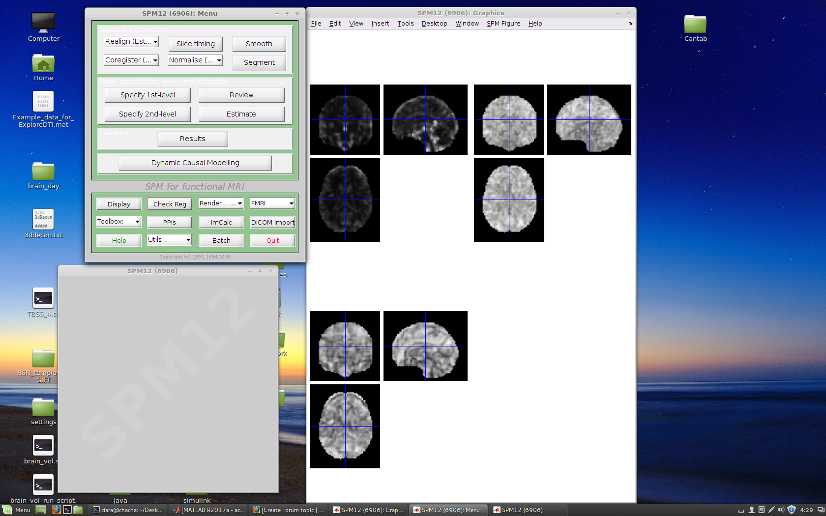
I carried out an analysis of the control group first and wanted results for ALFF, fALFF, reHO and FC, after DPARSF did all its processing, I was left with the results folder, that contained 4 folders for ALFF, fALFF, reHO and FC.
My FC folder is empty for some reason (anyone know why?). The files in my ALFF, fALFF and reHO should fmri in were black and white, which left me a bit confused. I assumed there would be coulours which indicated the different fluactions/activity of the brain. Is there another step I must carry out to carry out in order for colours to be seen? or Where ave I gone wrong.
Also can anyone suggest a manaual that goes through all the steps od DPARSF with a samaple R-fMRI data and explains how to intereperate thr results? The tutorial videos on this site, don't actaually show the process of getting results from the analysis.
I've also attached a screenshot, of my ALFF, fALFF and reHO results respectively, (should there be colour?)
regards..
| Attachment | Size |
|---|---|
| Screenshot from 2017-06-23 16:29:08.png | 1.01 MB |
| ALFF pic | 200.35 KB |
| Error image from matlab | 164.56 KB |
| DPARSF window | 198.28 KB |
Re: [RFMRI] What should my
Thank You for your reply,
Thank You for your reply,
So I think you are right, I didn't define a ROI for the FC section. How do I define a ROI? (we are looking at at the septal nuclei).
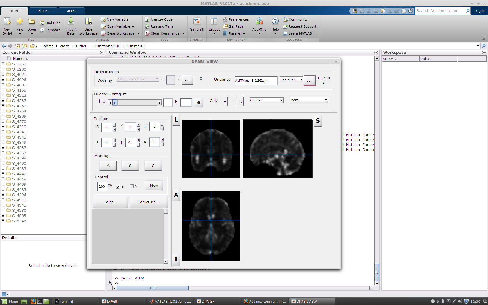
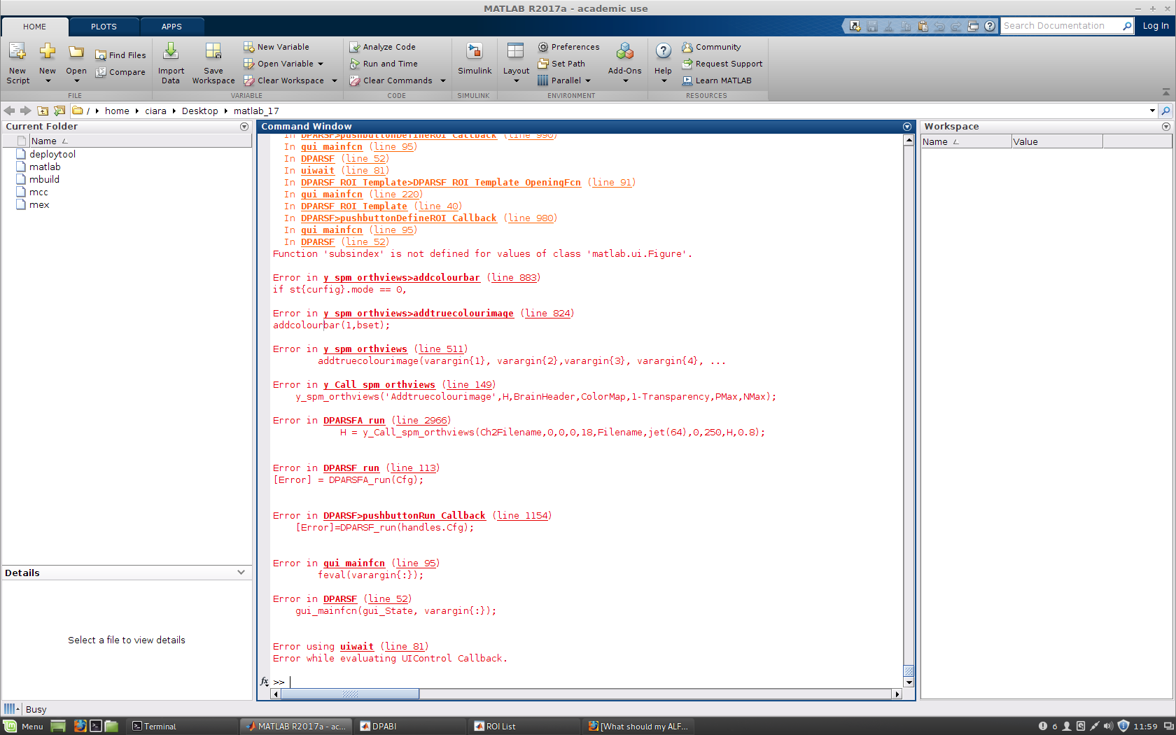
I viewed my ALFF and reHO results in DAPBI_VIEW and it is still colourless, there was an error message which came on matlab reagarding colour, I will add a screenshot of it.
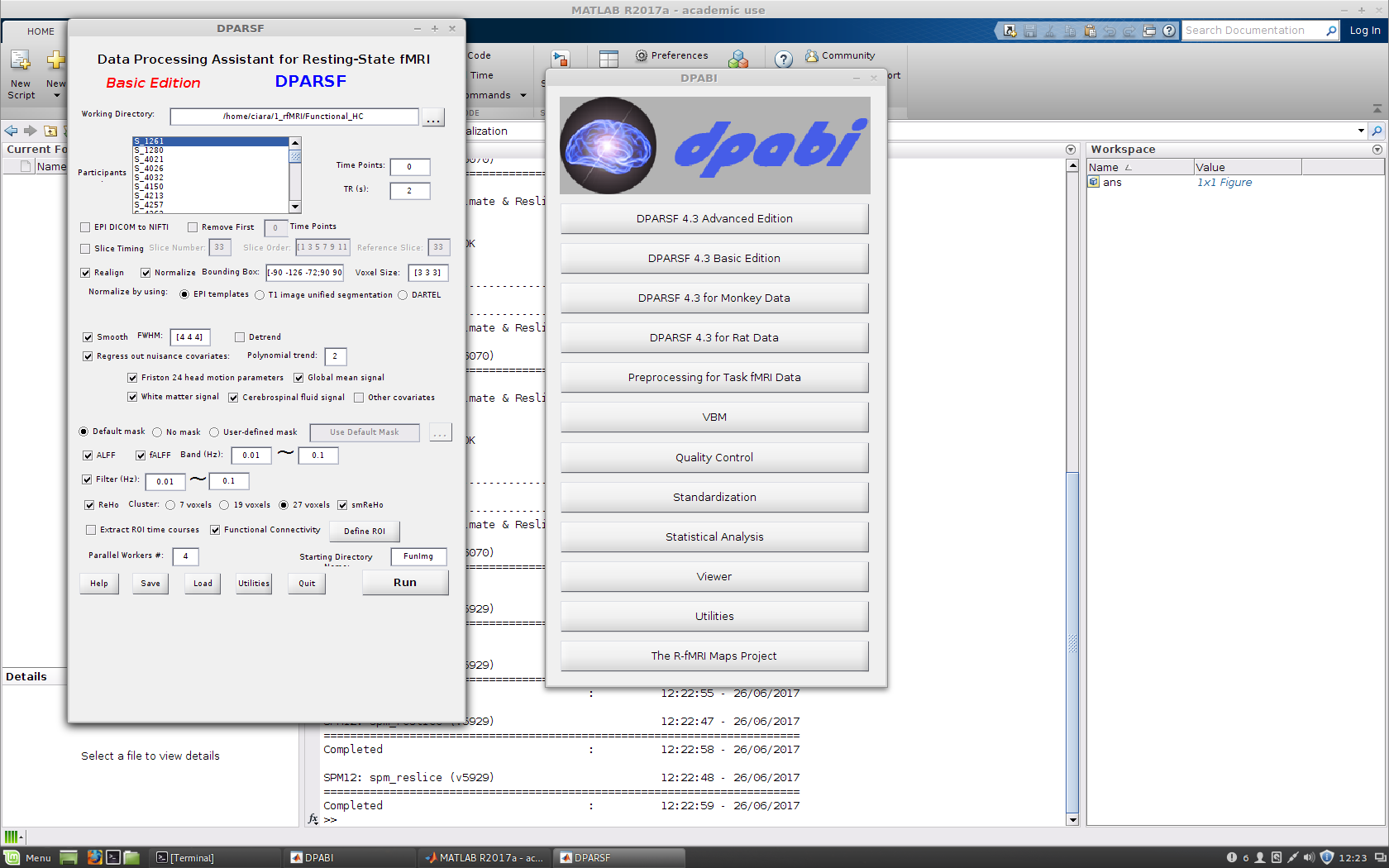
Also I will add a screenshot of what the ALFF image looks like as well as my DPARSF window.
(the screenshots are added to the initial post!)
Define ROI->Other ROIs-
Define ROI->Other ROIs->select the mask.
For the error message, you can try to restart matlab. If it still exists, it may be a problem of the comparability of matlab 2017.