blogs

  • 求助VMHC的一个问题

    Submitted by liufeng on
    Forums
    大家好,最近我打算用rest试着跑VMHC,出现以下问题:

    我直接打开rest的,然后输入数据目录,提示没数据。
    我不知道做VHMC需要哪步的数据,我试过这些目录的数据,都提示no data:
    FunImgNormalizedSmoothedDetrendedFiltered
    FunImgNormalizedSmoothedDetrendedFilteredCovremoved

    应该选择哪一步的数据呢?

    谢谢
  • 能否采用reho或alff进行纵向研究?

    Submitted by yxyxx111 on
    Forums
    大家好!向大家求教一个问题:能否采用reho或alff进行纵向研究? 我对同一批人间隔三年进行了两次resting-fmri的扫描,但是我不知道采用配对样本t检验对前后数据进行处理在方法学上是否行得通,因为fmri的可重复性并不是特别好。另外,如果可以的话,在数据处理时,除了采用配对样本t检验之外,还有没有其他的事情需要引起注意(就是和横断面研究有什么区别)?先谢谢大家了!
  • 请教严老师和大家!

    Submitted by will on
    Forums
    请教严老师和大家一个问题,我把20个患者和20个健康对照组的ReHo数据做了双样本t检验,使用AlphaSim校正,p值取0.05,但是结果显示双侧半卵圆中心脑白质大片脑活动增强的区域,不知道是怎么回事啊?迫切期待您们的帮助和解答!PS:只有一名患者头动超过1mm,其他的配合都很好。附图:图1是患者组单样本t检验的结果,图2是对照组单样本t检验的结果,图3是患者组-对照组双样本t检验结果。
  • Resting-State fMRI Data Analysis Toolkit V1.8

    Submitted by REST-Group on
    Forums
    Predefined Types

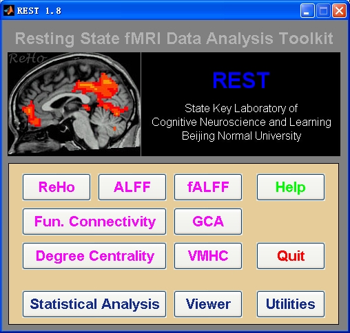
    Resting-State fMRI Data Analysis Toolkit (REST) is a convenient toolkit to calculate Functional Connectivity (FC), Regional Homogeneity (ReHo), Amplitude of Low-Frequency Fluctuation (ALFF), Fractional ALFF (fALFF), Gragner causality, degree centrality, voxel-mirrored homotopic connectivity (VMHC) and perform statistical analysis. You also can use REST to view your data, perform Monte Carlo simulation similar to AlphaSim in AFNI, perform Gaussian random field theory multiple comparison correction like easythresh in FSL, calculate your images, regress out covariates, extract ROI time courses, reslice images, and sort DICOM files. Download a MULTIMEDIA COURSE would be helpful for knowing more about how to use this software. Add REST's directory to MATLAB's path and enter "REST" in the command window of MATLAB to enjoy it.

    Citation of REST is: 
    Xiao-Wei Song, Zhang-Ye Dong, Xiang-Yu Long, Su-Fang Li, Xi-Nian Zuo, Chao-Zhe Zhu, Yong He, Chao-Gan Yan, Yu-Feng Zang. (2011) REST: A Toolkit for Resting-State Functional Magnetic Resonance Imaging Data Processing. PLoS ONE 6(9): e25031. doi:10.1371/journal.pone.0025031


    The latest release is
    REST_V1.8_130615


    DOWNLOAD 

    Multimedia Course: Data Processing of Resting-State fMRI

    New features of REST V1.8 release 130615:
    1. Fixed a bug in temporal correlation of two groups of images in Image Calculator. (Thanks for the report of ZHANG Han)

    2. The midline of VMHC results were set to zero. (YAN Chao-Gan)
     

    New features of REST V1.8 release 130303:
    When calling Mingrui Xia's BrainNet Viewer (http://www.nitrc.org/projects/bnv/), the default surface template is changed to the smoothed version (BrainMesh_ICBM152_smoothed.nv). The previous default template (BrainMesh_ICBM152.nv) hide more information in the sulcus. If the users want to use BrainMesh_ICBM152.nv as default surface template, please uncomment Line 3740 in rest_sliceviewer: %SurfFileName=[BrainNetViewerPath,filesep,'Data',filesep,'SurfTemplate',filesep,'BrainMesh_ICBM152.nv'];
    (After discussion with Mingrui Xia).

    New features of REST V1.8 release 130214:
    1.    This release fixed some minor bugs, will not affect any data analysis.
    2.    Fixed a bug when using .nii(.gz) files in REST Image Calculator. (WANG Xin-Di)
    3.    Fixed a bug in using .nii(.gz) files in GCA analyses. (ZANG Zhen-Xiang)
    4.    Fixed the imresize_old bug of REST Slice Viewer with Matlab 2012b. (YAN Chao-Gan)

    New features of REST V1.8 release 121225:
    1.    Support parallel computing! If you installed the MATLAB parallel computing toolbox, REST can distribute the subjects into different CPU cores. (WANG Xin-Di and YAN Chao-Gan).
    2.    Algorithm change: (1) Filtering: a separate function for matrix filtering was written. The low cutoff frequency index calculation changed from round (in REST V1.7) to "ceil". E.g., if low cut off corresponded to index 5.1, now it will start from 6 other than 5. This change also applies to ALFF and fALFF calculation. The filtered data changes slightly, about 0.0001. (2) The ALFF generated by the new version is sqrt(2/N) times of the original version. (new version used: 2*abs(fft(x))/N; original version used:  sqrt(2*abs(fft(x))^2/N)). This change will not affect group analysis (as each individual scaled the same number), and will not affect mALFF and fALFF calculation as this factor will be normalized. (3) In the calculation of ReHo, the rank will keep as double and no longer converted into uint16, thus created slight difference with REST V1.7. (YAN Chao-Gan)
    3.    REST Slice Viewer support 4D file display and the maximum and minimum value could be set. (WANG Xin-Di)
    4.    Gaussian random field (GRF) theory multiple comparison correction (like easythresh in FSL) was supported. The smoothness could be evaluated for GRF correction or AlphaSim correction. (GUI by WANG Xin-Di, algorithm by YAN Chao-Gan)
    5.    Modules of voxel-mirrored homotopic connectivity (VMHC) (Zuo et al., 2010), Degree Centrality (Buckner et al., 2009) were added. (GUI by WANG Xin-Di, algorithm by YAN Chao-Gan)
    6.    REST GCA: could handle multiple ROIs (other than 2) in ROI-wise GCA now. Fixed a bug of discordance between the outputs and the description in REST-GCA readme in the pre-release of REST V1.8. (ZANG Zhen-Xiang)
    7.    rest_readfile.m and rest_writefile: The default format changed to .nii from .img. (WANG Xin-Di)
    8.    rest_to4d.m: now support one 4d file other than a directory, also support a cell of image filenames. (YAN Chao-Gan)
    9.    rest_regress_ss.m: add the output of T value. (YAN Chao-Gan)
    10.    rest_Write4DNIfTI.m: This function was added for write 4D nifti files based on SPM’s nifti function. (YAN Chao-Gan)
    11.    rest_writefile.m: No longer need to change to RPI before writing. (YAN Chao-Gan)

  • Data Processing Assistant for Resting-State fMRI (DPARSF) V2.2

    Submitted by YAN Chao-Gan on
    Forums
    Predefined Types

    Data Processing Assistant for Resting-State fMRI (DPARSF) is a convenient plug-in software based on SPM and REST. You just need to arrange your DICOM files, and click a few buttons to set parameters, DPARSF will then give all the preprocessed (slice timing, realign, normalize, smooth) data, functional connectivity, ReHo, ALFF/fALFF, degree centrality, voxel-mirrored homotopic connectivity (VMHC) results. DPARSF can also create a report for excluding subjects with excessive head motion and generate a set of pictures for easily checking the effect of normalization. You can use DPARSF to extract ROI time courses efficiently if you want to perform small-world analysis. DPARSF basic edition is very easy to use while DPARSF advanced edition (alias: DPARSFA) is much more flexible and powerful. DPARSFA can parallel the computation for each subject, and can be used to reorient your images interactively or define regions of interest interactively. You can skip or combine the processing steps in DPARSF advanced edition freely. Please download a MULTIMEDIA COURSE to know more about how to use this software. Add DPARSF's directory to MATLAB's path and enter "DPARSF" or "DPARSFA" in the command window to enjoy DPARSF basic edition or advanced edition.

    The latest release is DPARSF_V2.2_130309

    DOWNLOAD 

    Multimedia Course: Data Processing of Resting-State fMRI
    New features of DPARSF_V2.2_130309:
    1. Advanced Edition: Fixed a bug in "Smooth by DARTEL" caused in previous revision (DPARSF_V2.2_130214). (Thanks for the report of Maki Koyama).

    New features of DPARSF_V2.2_130303:
    1. Advanced Edition: Fixed a bug in processing of multiple sessions - only process the last session in normalization and smooth.
    2. Basic Edition: Fixed a bug caused in the previous release (V2.2_130214) - cannot load the correct masks (Thanks for the report of Tao Yang).

    New features of DPARSF_V2.2_130224:
    1.    This release fixed some minor bugs, but will not affect the results of any data analysis. The bugs appear in uncommon parameter settings and stop the processing in the worst cases.
    2.    Updated the DPARSFVersion information in the template parameter files.
    3.    DPARSF basic edition will also output the results of ReHo/ALFF/fALFF after Z-standardization (subtract the whole brain mean and divide by the whole brain standard deviation).
    4.    Fixed a bug in nuisance covariates regression when CovMat is not defined.
    5.    Fixed a bug that can not save the ROI signals correctly in text file if the data is not in double format. (Thanks for the report by H. Baetschmann)
    6.    Fixed a bug in creating mean functional image for 4D files. (Thanks for the report and revision by S. Orsolini)
    7.    Fixed a bug in displaying ROI Templates in linux system. (Thanks for the report by Han Zhang)

    New features of DPARSF_V2.2_121225:
    1.    Support parallel computing! If you installed the MATLAB parallel computing toolbox, you can set the number of "Parallel Workers", DPARSFA will distribute the subjects into different CPU cores.
    2.    In addressing head motion concerns in resting-state fMRI analyses (Power et al., 2012; Satterthwaite et al., 2012; Van Dijk et al., 2012), we provide Friston 24-parameter correction (See Yan et al., 2013 for a comprehensive assessment of head motion on functional connectomics) as well as voxel-specific head motion calculation and correction (Satterthwaite et al., 2013; Yan et al., 2013). DPARSF also calculate the voxel-specific mean framewise displacement (FD) and volume-level mean FD (Power) (Power et al., 2012) or FD (Jenkinson) (i.e., relative RMS; Jenkinson et al., 2002) for accounting head motion at group-level analysis. The data scrubbing approach is also supported with different methods: 1) model each bad time point as a separate regressor in nuisance covariates regression, 2) delete bad time points, 3) interpolate bad time points with nearest neighbor, linear or cubic spline interpolation. 
    3.    According to (Weissenbacher et al., 2009), the nuisance covariate regression could be performed before filtering and at very early stage. Users can also choose Template Parameters: TRADITIONAL order to have the same order as the previous version.
    4.    Support .nii.gz in all the steps. No longer need to convert 4D .nii.gz into 3D .img/.hdr. Simply put .nii.gz under FunImg or any starting directory name, DPARSFA will handle the .nii.gz by itself.
    5.    If the Number of Time Points is set to 0, then DPARSFA will not check the number of time points.
    6.    If TR is set to 0, then DPARSFA will retrieve the TR information from the NIfTI images. Please ensure the TR information in NIfTI images are correct!
    7.    If Slice Number is set to 0, then retrieve the slice number from the NIfTI images. The slice order is then assumed as interleaved scanning: [1:2:SliceNumber, 2:2:SliceNumber]. The reference slice is set to the slice acquired at the middle time point, i.e., ceil(SliceNumber/2). SHOULD BE EXTREMELY CAUTIOUS!!!
    8.    Support calculating resting-state fMRI metrics in native space and warping by DARTEL. The mask files and/or region of interest (ROI) files in standard space are warped into native space by using the parameters estimated in segmentation or DARTEL.
    9.    Spatial normalization and smooth can be performed on the calculated resting-state fMRI derivatives.
    10.    Supporting extracting ROI time course using masks with multiple labels. More template ROI definitions are supported: Dosenbach’s 160 functional ROIs (Dosenbach et al., 2010), Andrews-Hanna’s default mode network ROIs (Andrews-Hanna et al., 2010), Craddock’s clustering ROIs (Craddock et al., 2011), AAL atlas and Harvard-Oxford atlas.
    11.    More resting-state fMRI metrics are included, e.g., voxel-mirrored homotopic connectivity (VMHC) (Zuo et al., 2010), Degree Centrality (Buckner et al., 2009) and connectome-wide association studies based on multivariate distance matrix regression (Shehzad et al., 2011).
    12.    For VMHC analyses, support normalizing the data further to a symmetric template. 1) Get the T1 images in MNI space (e.g., wco*.img or wco*.nii under T1ImgNewSegment or T1ImgSegment) for each subject, and then create a mean T1 image template (averaged across all the subjects). 2) Create a symmetric T1 template by averaging the mean T1 template (created in Step 1) with it's flipped version (flipped over x axis). 3) Normalize the T1 image in MNI space (e.g., wco*.img or wco*.nii under T1ImgNewSegment or T1ImgSegment) for each subject to the symmetric T1 template (created in Step 2), and apply the transformations to the functional data (which have been normalized to MNI space beforehand). Please see a reference from Zuo et al., 2010.
    13.    A group analyses function was added as y_GroupAnalysis_Image by scripting call. T tests or F tests could be performed for a given set of regressors.
    14.    Template Parameters:
             Calculate in Original Space (warp by DARTEL)
             Calculate in Original Space (warp by information from unified segmentation)
             Calculate in MNI Space (warp by DARTEL)
             Calculate in MNI Space (warp by information from unified segmentation)
             Calculate in MNI Space: TRADITIONAL order [This is the order used in DPARSF basie edition as well as DPARSFA V2.1]
             Intraoperative Processing
             Task fMRI data preprocessing
             VBM (New Segment and DARTEL)
             VBM (unified segmentation)
             Blank

    Many thanks to Dr. Chris Rorden for suggesting features 4-7. Many thanks to Dr. Susan Whitfield-Gabrieli for discussing the head motion scrubbing regressors.

  • 我做双样本T检验的mask做法对吗,谢谢!!!

    Submitted by shuxie on
    Forums
    各位老师: 我想咨询一下双样本T检验的mask问题, 我是MRI新手,我的感受是Rest软件最大的困难对我来说是每一个统计都让你选mask,而我不会制作mask,不点mask总觉得分析不规范,为此我为了mask摸索了一个月, 才敢用此软件。而SPM的分析只是点击,选默认mask。我根据严老师讲课视频和论坛中有老师写的"先用两组单样本T检验显著的脑区,在RESTimage Calculator:中根据公式(abs(i1)> threshold)+(abs(i2)> threshold)>0取他们的并集做一个mask供双样本检验用,这样小一点的mask才有可能使有些信息活下来的"这个原则,我进行了mask制作.但我不知道我做得对不对: 1. 我用DPARS算出两组各个人的smRehoMap等, 先对两组分别进行smReho-1 MAP单样本的T检验,因我没有假设,所以这步mask 我选的Rest软件自带的brain61X72X61模板作为mask(因我看文章中阳性区域报的结果太多了,尽管基底节更多一些), 然后, 单样本T检验中得到病人和对照组各自的T图,下一步,取两组T检验显著的脑区,先去看threshold value, 用RestSlice Viewer中将病人T图呈现出来, 我选择P=0.05, 得出Threshold=2.1448,又将对照组T图呈现出来, 选择P=0.05,得出Thresthold=2.093.有了threshold, 可以在Rest Image Calculator中根据公式: (abs(i1)> threshold)+(abs(i2)> threshold)>0, 输入(abs(i1)> 2.1448)+(abs(i2)> 2.093)>0, 出来的mask是个红色图(见图1,光盘映像文件,需用Rest Sliceview打开),我不知道这个过程或公式对否, abs是什么意思,是否不要加? 2. 于是, 我去掉abs, 又用(i1> 2.1448)+(i2> 2.093)>0(我看视频上为(i1>1.96+i2>1.96)>0)公式,出来的图比刚才那个图少些脑组织(见图2),不知道哪个对? 3. 最后我用第一个公式做出来的mask,作双样本T检验, 结果统计出来的下面的T图(见图3),但不知道是什么意思,这是 smReho 最后分析出来的是结果吗,我选P=0.05,cluster size=85, rmm=4,选了个图像裁了下来(见图4)? 4. 另外一个问题是如果不用大脑或灰质模板做mask的话,是否每做一个变量都要做mask,如分析mReho,ALFF, mALFF,fALFF等要按上述方法制作阳性结果并集的mask吗? 请各位能给予回复和指导,谢谢~~~~