| Attachment | Size |
|---|---|
| DPARSF_FC_subject1and2.jpg | 142.55 KB |
Dear all
Hi,
I'm using DPARSF for 12 subjects.
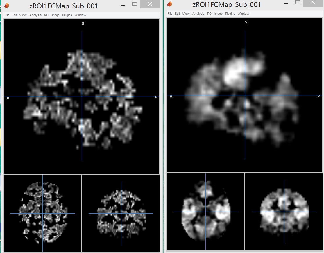
Today I found that in one subject, functional connectivity images from all 4 sessions of BOLD resting state fMRI have different image from others.
Here I attached a screenshot, left one is different from right normal image.
I did every analysis in same procedure and checked several times.
It seems like participants moved during measurement, but when I checked a realignment parameter, movement is less than 0.5mm.
T1 Image of this participant seems that he may move during T1 imaging.
Is this could be a reason?
If I measure T1 again, do you think this problem could be solved?
Thanks
Bests,
Inseon
Probably just exclude this
Probably just exclude this subject.